Modeling Principle
The Mouse U-87 MG Model is an in-vivo tumor model established by subcutaneous inoculation or orthotopic intracranial inoculation of the U87MG human glioblastoma cell line into immunodeficient mice (BALB/c nude mice or NOD/SCID mice), serving as one of the most widely applied and most frequently reported standardized in-vivo gold-standard tumor models in glioma translational medicine, antitumor drug pharmacodynamics, radio/chemosensitization, targeted therapy and tumor biology mechanism research. Immunodeficient mice lack mature T cells (nude mice) or combined T/B/NK immunodeficiency (NOD/SCID), allowing xenogeneic transplantation of human U87MG cells without immune rejection. Tumor cells proliferate stably in mice to form solid tumors highly consistent with human glioblastoma histology: high cellular density, marked nuclear atypia, microvascular proliferation, pseudopalisading necrosis and surrounding tissue infiltration as typical GBM pathological structures. As one of the longest-established glioblastoma cell lines, U87MG features mature culture protocols, stable batches, high take rate, rapid growth and short doubling time (approximately 24–30 h), with stable xenograft phenotype and highly reproducible tumor growth kinetics and volume/weight among individuals, serving as the most widely used cell line model in global glioma research. The subcutaneous model facilitates dynamic tumor volume measurement, diverse administration routes and repeated sampling, suitable for standard pharmacodynamic evaluation and histopathological analysis. The orthotopic intracranial model more closely mimics the true anatomical location and microenvironment of human glioma, allowing evaluation of invasive tumor growth, neurological symptoms and survival. The U87MG xenograft model is widely applied in anti-glioma drug screening, mechanism validation, radio/chemosensitization, resistance reversal, nano-delivery and imaging monitoring (MRI, bioluminescence imaging BLI) studies, acting as an indispensable in-vivo gold-standard carrier for preclinical glioma research.
Model Validation Criteria
Quantitative Criteria for Tumor Formation and Growth
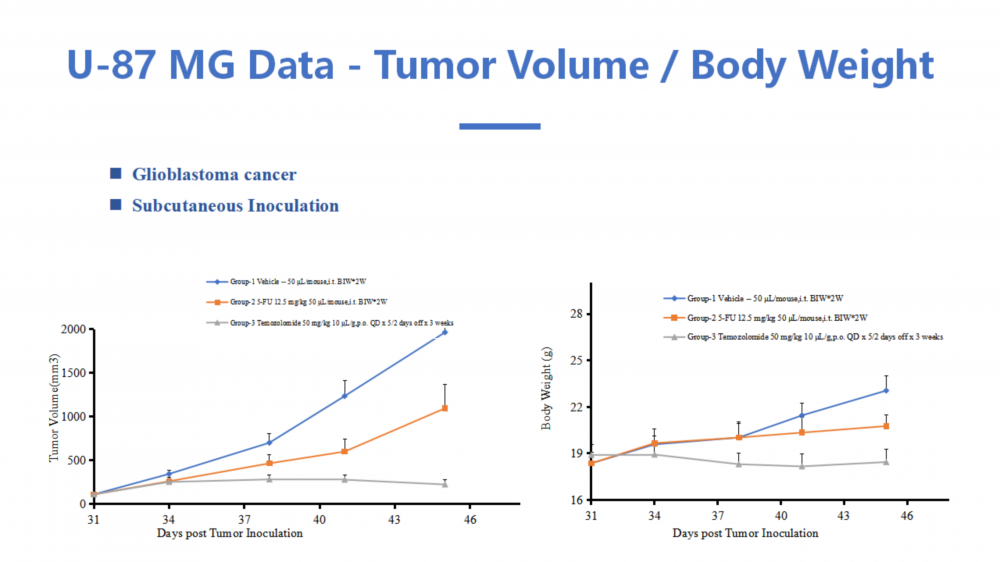
1. Tumor take rate: subcutaneous model achieves ≥95% take rate within 2–3 weeks after inoculation (tumor volume >50 mm³ judged as successful tumor formation), orthotopic model confirmed by pathological evidence of intracranial tumor; 2. Tumor growth: vehicle control group shows stable, sustained and reproducible growth, with average tumor volume reaching 800–1500 mm³ at weeks 3–4 after inoculation; 3. Inter-group consistency: coefficient of variation (CV) of tumor volume in vehicle control group <25%, confirming excellent inoculation operation and model reproducibility; 4. Positive drug validation: TGI of TMZ positive control group ≥40%, confirming sensitive and effective pharmacodynamic evaluation system, and the model is judged successfully established for stable drug screening.
Histopathological and Immunohistochemical Gold-Standard Indexes
1. HE staining: xenografts present typical histological features of human glioblastoma—high cellular density, marked nuclear atypia, microvascular proliferation, pseudopalisading necrosis and brain tissue infiltration (orthotopic model); 2. Ki-67 proliferation index: Ki-67 positive rate of treatment group significantly decreased compared with vehicle group (P<0.05), indicating proliferation suppression; 3. TUNEL apoptosis: significantly increased apoptotic cells in treatment group, confirming pro-apoptotic effect; 4. CD31 microvessel density (MVD): MVD of treatment group decreased compared with vehicle group, indicating anti-angiogenic effect (optional); 5. Molecular indexes: down-regulation of proliferation-, invasion- and resistance-related proteins and up-regulation of apoptotic proteins in treatment group, cross-validated with in-vitro mechanism results, confirming closed-loop efficacy and mechanism.
Academic Evidence for Model Validity
Core validation evidence for U87MG xenograft model: ① immunodeficient mice successfully tolerate xenogeneic transplantation of human U87MG cells with high take rate and stable tumor phenotype; ② xenograft histology highly consistent with human GBM; ③ reproducible tumor growth with controllable individual variation meeting pharmacodynamic statistical requirements; ④ positive drug control system validated effective, confirming model sensitivity to drug intervention; ⑤ orthotopic model evaluates invasive growth, survival and neurological function, closely mimicking clinical glioma course. Combined verification of above indexes confirms model reliability and reproducibility, serving as the most widely applied gold-standard model carrier for in-vivo glioma efficacy and mechanism research.
Model Advantages
The U87MG xenograft model is the most frequently reported in-vivo glioma model in literature, featuring mature cell culture, stable batches, high take rate (≥95%), rapid growth and short experimental cycle. Immunodeficient mice completely retain the malignant phenotype and histological features of human GBM with high clinical translational value. Subcutaneous model features convenient operation, non-invasive dynamic tumor volume measurement, diverse administration routes and repeated sampling, suitable for standard pharmacodynamic evaluation. Orthotopic model closely mimics the anatomical location and microenvironment of human glioma, enabling evaluation of invasive growth, survival and neurological function. U87MG cells possess defined background with reproducible xenograft phenotype and low intra-group variation, offering the strongest cross-study data comparability in global glioma research. It supports diverse research designs including monotherapy/combination sensitization, radiosensitization, resistance reversal, nano-delivery and imaging monitoring. Complete detection system (tumor growth + pathology + immunohistochemistry + molecular mechanism + toxicological safety) forms closed-loop evidence chain, and results are readily accepted by neuro-oncology and pharmacology SCI journals, suitable for NSFC projects, postgraduate dissertations and research proposals.
Research Applications
The Mouse U-87 MG Model is mainly applied to evaluate in-vivo antitumor efficacy, dose-effect relationship and safety of anti-glioma drugs, natural active products, nano-delivery systems, gene therapy and radio/chemosensitization strategies; to dissect in-vivo regulatory mechanisms of glioma proliferation, invasion, apoptosis, resistance, angiogenesis and signaling pathways; to establish TMZ-resistant xenograft models for resistance reversal and mechanism research; to perform imaging (BLI/MRI)-guided dynamic tumor monitoring and efficacy evaluation; to optimize drug combination regimens and translational medical validation. It extensively serves basic and clinical translational research on glioma, targeted new drug development, NSFC projects, postgraduate dissertations and SCI manuscript methodology construction, acting as the standardized in-vivo gold-standard model with the largest literature volume and widest application in neuro-oncology.
U87MG glioblastoma nude mouse subcutaneous xenograft model, orthotopic intracranial glioma model, GBM xenograft tumor, in-vivo antitumor efficacy evaluation, temozolomide resistance, tumor growth inhibition rate, survival analysis, in-vivo tumor pharmacology
 Zvast Biotechnology