

Hope for Millions! Disruptive Breakthroughs in Cerebral Hemorrhage Treatment—Innovative Approaches Rewrite Prognosis
One in three stroke patients dies or is left permanently disabled due to intracerebral hemorrhage (ICH)! As the subtype of stroke with the highest mortality and disability rates, ICH affects over one million new patients annually in China, with an acute-phase mortality rate of 30%–40%. Nearly 70% of survivors suffer from sequelae such as paralysis and cognitive impairment.
For a long time, clinical treatment has been mired in the dilemma of symptomatic but non-targeted therapy. In recent years, however, breakthroughs in neuroprotection, precision minimally invasive procedures and other fields are reshaping this landscape, bringing new hope for rehabilitation to patients.
I. Etiology of Intracerebral Hemorrhage
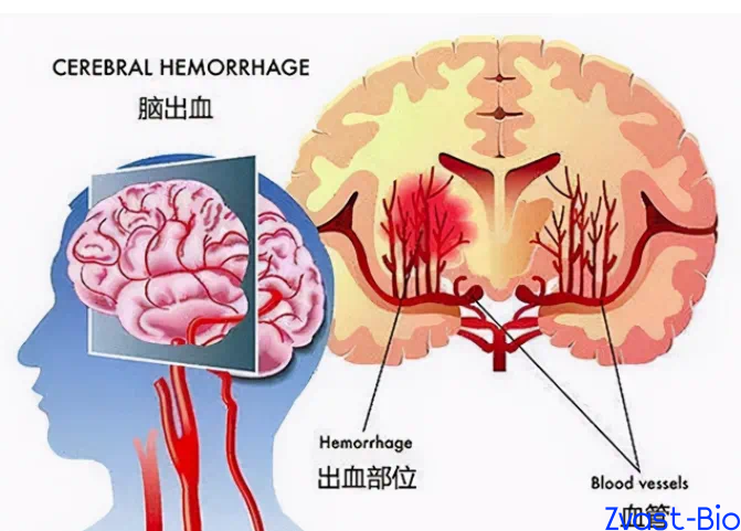
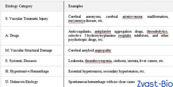
Intracerebral hemorrhage (ICH) refers to non-traumatic vascular rupture and hemorrhage within the brain parenchyma. Hypertension complicated with arteriolosclerosis is the predominant etiology, accounting for over 60% of cases.
Most patients develop symptoms during physical activity or emotional agitation, with typical manifestations including sudden headache, nausea and vomiting, limb paralysis, and disturbance of consciousness. The specific clinical presentation is closely correlated with the hemorrhage location: putaminal hemorrhage is the most common type, which tends to cause contralateral hemiplegia; thalamic hemorrhage is characterized by prominent sensory disturbance; brainstem or cerebellar hemorrhage progresses rapidly, often accompanied by respiratory and circulatory dysfunction, with an extremely high mortality rate.
II. Global Treatment Hotspots: From Symptomatic Intervention to Targeted Repair
To address key clinical challenges, research teams worldwide are breaking through therapeutic bottlenecks across multiple dimensions, forming a diversified treatment paradigm featuring precision minimally invasive evacuation, targeted neuroprotection, and endogenous regenerative repair.
Sustained increases in R&D investment have served as a critical driver of innovative breakthroughs. From 2023 to 2025, the global R&D expenditure on innovative drugs for intracerebral hemorrhage has witnessed an average annual growth rate of 22%. Neuroprotection and anti-edema have emerged as core R&D focus areas, with multiple drug candidates advancing to pivotal development phases.
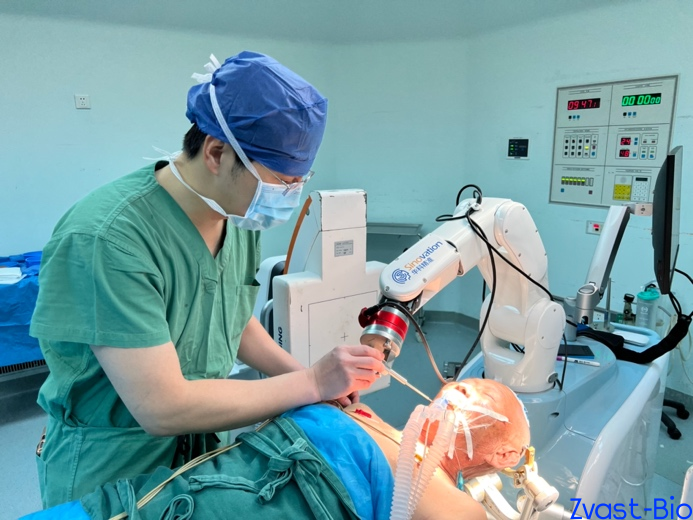
1. Precision Minimally Invasive Evacuation: Farewell to Craniotomy, the "Chinese Solution" Shines
Traditional craniotomy for hematoma evacuation is highly invasive with significant brain tissue traction, only suitable for critically ill patients with massive hemorrhage. In recent years, precision minimally invasive technologies centered on neuroendoscopy and stereotactic puncture drainage have become mainstream. Combined with hematoma liquefiers, these technologies enable hematoma evacuation with minimal trauma, significantly reducing the risk of complications such as rebleeding and infection.
Precision minimally invasive technology has now become the primary direction for hematoma evacuation, with sustained vitality in technological innovation. According to database statistics, the global compound annual growth rate of patent applications for neuroendoscopy, robot-assisted surgical instruments and related devices has reached 18% in the past five years.
Domestically, clinical breakthroughs are remarkable. Dezhou Hospital of Qilu Hospital of Shandong University has launched a multicenter clinical trial to verify the efficacy of the combined regimen of "neuro-navigation assisted stereotactic minimally invasive puncture + tenecteplase for hematoma lysis" . Focusing on the precise treatment of deep intracerebral hemorrhage, this regimen is expected to form an internationally influential "Chinese Solution" .
Internationally, robot-assisted puncture technology achieves sub-1mm precise positioning through AI algorithms , greatly improving the safety of deep hemorrhage treatment. Postoperatively, patients show significantly reduced neurological deficit scores, and the rebleeding rate is controlled below 5%.
2. Emergence of Innovative Neuroprotective Drugs: Solving the Problem of Secondary Injury
Secondary injury is the core cause of poor prognosis in patients with intracerebral hemorrhage (ICH). How to simultaneously inhibit oxidative stress and inflammatory responses, as well as promote nerve regeneration, has become the core goal of current drug research and development.
The global R&D pipeline of innovative drugs for ICH has exceeded 40 candidates, among which neuroprotection and anti-edema candidates account for the highest proportion and have become R&D hotspots.
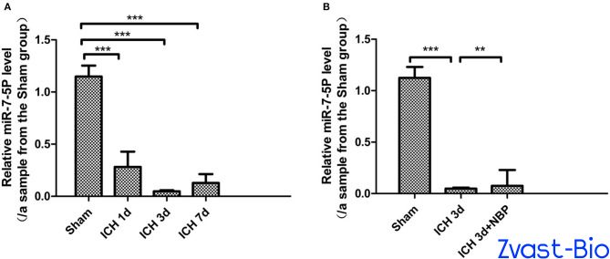
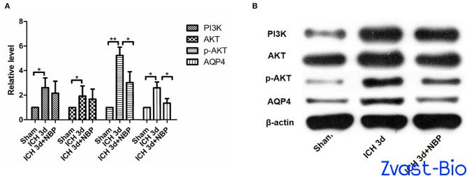
Domestic scholar teams have achieved significant breakthroughs in research and found that butylphthalide has shown outstanding performance in improving the therapeutic effect of ICH. Studies have confirmed that butylphthalide can up-regulate the levels of STAT3 protein and miR-7-5p, inhibit the EGFR/PI3K/AKT pathway and AQP4 expression, fundamentally reduce the occurrence of cerebral edema, block inflammation and cellular edema pathways, and lower the risk of secondary brain injury.
Similarly, the injectable biomimetic hydrogel developed by the team of West China Hospital deserves attention. It has the functions of scavenging reactive oxygen species, inhibiting inflammation and targeted drug delivery, and can promote the proliferation and differentiation of neural stem cells as well as angiogenesis. At present, it has entered the preclinical transformation stage.

3. AI-Powered New Drug R&D: Opening Up a Brand-New Track
The global innovative drug R&D landscape also features remarkable highlights. Beyond the exploration of targeted neuroprotectants, AI-powered new drug R&D has emerged as a brand-new track.
As reported, Qanatpharma (Switzerland), in collaboration with institutions including the Zuse Institute Berlin, has launched a generative AI-driven R&D project focusing on delayed cerebral ischemia (DCI) complications induced by subarachnoid hemorrhage. This project integrates the entire workflow spanning target validation, molecular design, compound synthesis, and structural verification. It leverages generative algorithms to rapidly screen candidate molecules, and combines high-precision structural analysis technology to optimize drug design. Expected to break through the limitations of existing therapies, this initiative is poised to provide a groundbreaking treatment regimen for approximately 500,000 related patients worldwide, with preclinical research phases scheduled to commence in 2026.
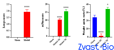
For neurologists and pharmaceutical R&D professionals, these key focus areas present both challenges and opportunities. Currently, innovative drug R&D for intracerebral hemorrhage still faces bottlenecks such as difficulties in mechanism validation and low efficacy translation efficiency. High-quality preclinical research serves as the key to overcoming these hurdles, among which animal models accurately matching the pathological characteristics of the disease act as the core bridge connecting basic research and clinical application.
For the general public, controlling hypertension, quitting smoking and limiting alcohol intake, and maintaining a regular daily routine are the most effective measures to prevent intracerebral hemorrhage. In the event of suspected symptoms such as sudden headache or limb numbness, immediate medical attention is imperative to secure valuable time for treatment.
References
Chen X, Deng S, Lei Q, He Q, Ren Y, Zhang Y, Nie J, Lu W. miR-7-5p Affects Brain Edema After Intracerebral Hemorrhage and Its Possible Mechanism. Front Cell Dev Biol. 2020 Dec 16;8:598020. doi: 10.3389/fcell.2020.598020. PMID: 33392188; PMCID: PMC7772315.
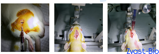
Autologous Blood-Induced Intracerebral Hemorrhage (ICH) Model in SD Rats
Experimental Animals: Male SD rats, 8 weeks old
2. Preoperative Preparation: Anesthetization, scalp shaving and disinfection
3. Grouping Procedure
◦ Sham-operated Group: The scalp was incised and then sutured directly.
◦ Model Group: A midline incision was made on the scalp; a burr hole was drilled at a specific location on the skull. Non-anticoagulated blood was collected from the caudal artery using a microsyringe and slowly injected into the brain in two separate aliquots, with the needle retained for a period after each injection. After needle withdrawal, the burr hole was sealed with paraffin. The scalp was sutured after confirming no hemorrhage, and the tail incision was compressed and bandaged.
Collagenase‑Induced Intracerebral Hemorrhage Model in SD Rats
Experimental Animals: Male Sprague-Dawley (SD) rats, 8 weeks old, were randomly grouped after adaptive feeding.
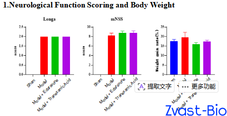
Grouping: Control group; Sham-operated group; Model group; Model + Edaravone group; Model + Tranexamic Acid group
Modeling Procedure: Referring to the brain stereotaxic atlas, a burr hole was drilled to localize the right striatum of rats, and collagenase solution prepared with normal saline was slowly injected via a microsyringe.
Positive Drugs: Edaravone; Tranexamic Acid
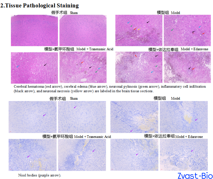
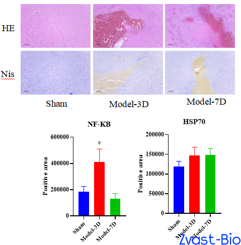
HE Staining Results
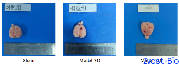
No obvious pathological changes were observed in the Sham Operation Group.
In all brain tissue samples of the Model Group, obvious hematoma was observed; around the hematoma, neuronal pyknosis or edema was seen, accompanied by inflammatory cell infiltration, and obvious glial cell proliferation was observed in a few samples.
In most brain tissue samples of the Model + Edaravone Group, obvious hematoma was observed; around the hematoma, neuronal pyknosis and edema were seen, accompanied by inflammatory cell infiltration, and glial cell proliferation was observed in a small number of samples.
In most brain tissue samples of the Model + Tranexamic Acid Group, obvious hematoma was observed; around the hematoma, neuronal pyknosis and edema were seen, accompanied by inflammatory cell infiltration.
Nissl Staining Results
In the Sham Operation Group, the morphology of Nissl bodies and neurons was normal, with no obvious pathological changes observed.
In all samples of the Model Group, some Nissl bodies showed elongated morphology or necrosis, and the number of Nissl bodies was significantly reduced; in some samples, the number of neurons around the hematoma was significantly decreased.
In most samples of the Model + Edaravone Group, some Nissl bodies showed elongated morphology or necrosis, and the number of Nissl bodies was significantly reduced; in a small number of samples, the number of neurons around the hematoma was significantly decreased.
In most samples of the Model + Tranexamic Acid Group, some Nissl bodies showed elongated morphology or necrosis, and the number of Nissl bodies was significantly reduced; in a small number of samples, the number of neurons around the hematoma was significantly decreased.